Nursing Pathophysiology Memory NoteCards: Hypovolemic Shock

Nursing Pathophysiology Memory NoteCards: Hypovolemic Shock

Hypovolemic Shock Nursing, Treatment, Management, Interventions NCLEX

Cardiogenic Shock Nursing Management, Pathophysiology, Interventions NCLEX Review

Hypovolemic Shock Nursing Care Plan

Hypovolemic shock happens when a sudden and significant loss of blood or body fluids drops your blood volume.

Treatment goals: fluid resuscitation, correct underlying cause that is leading to the fluid loss….example: hemorrhaging: surgery (get the patient ready for surgery).

Nursing Pathophysiology Memory NoteCards: Hypovolemic Shock

Tipos de Patrones Respiratorios: PATRONES RESPIRATORIOS ANORMALES

Tipos de Patrones Respiratorios: PATRONES RESPIRATORIOS ANORMALES

El control de la respiración se realiza en el tronco del encéfalo. Los patrones respiratorios pueden ayudar a localizar la zona de la lesión e incluso a sospechar la etiología:
– Apnea de posthiperventilación: sólo se explora en sujetos obnubilados, ya que requiere un mínimo de colaboración. Puede considerarse signo precoz de afectación de la conciencia. Tras solicitar al paciente que realice cinco inspiraciones profundas, los pacientes con alteración del sistema límbico presentan una pausa de posthiperventlación de 10 segundos, mientras que los sujetos sanos continúan respirando sin interrupciones.
– Respiración de Cheyne-Stokes: respiración periódica con alternancia de fases de aumento y descenso progresivo de frecuencia e intensidad respiratoria, seguido de un período de apnea de duración variable. Se observa en lesiones hemisféricas bilaterales, fase diencefálica del coma o encefalopatía metabólica. Existe otro tipo de patrón respiratorio similar al de Cheyne-Stokes, pero con ciclos de mucha menor duración, que ocurre en casos de hipertensión intracraneal grave y parece ser secundario a isquemia de tronco.
– Coma con hiperventilación: la hiperventilación es frecuente en el coma de origen metabólico, se asocia a acidosis metabólica: uremia, cetoacidosis diabética, ingesta de ácidos orgánicos, etc. o a alcalosis respiratoria (encefalopatía hepática e hipoxia de cualquier etiología).
– La hiperventilación neurógena central: es infrecuente y se caracteriza por una respiración continua, profunda y rápida, a una frecuencia de 25 ciclos por minuto. Se asocia a lesiones de mesencéfalo inferior y tercio medio de la protuberancia. Su diagnóstico obliga a excluir otras causas acidosis metabólica.
– Respiración apnéustica: signo muy específico de afectación de protuberancia inferior. Caracterizado por una bradipnea de fondo con períodos de apnea que pueden alcanzar los 30s, seguida de una inspiración profunda, tras la cual se detiene la respiración durante 10-15 segundos.
– Respiración atáxica: patrón respiratorio irregular en frecuencia y profundidad. Se observa en las lesiones de la región dorsomedial del bulbo. Indicador de mal pronóstico.

Tipos de Patrones Respiratorios: PATRONES RESPIRATORIOS ANORMALES

Immune System Overview for Nursing Students (Structure and Function)

Immune System Overview for Nursing Students (Structure and Function)

Types of immune responses: Innate and adaptive, humoral vs. cell-mediated

Innate vs. adaptive immunity. Humoral adaptive immunity vs. cell-mediated adaptive immunity.

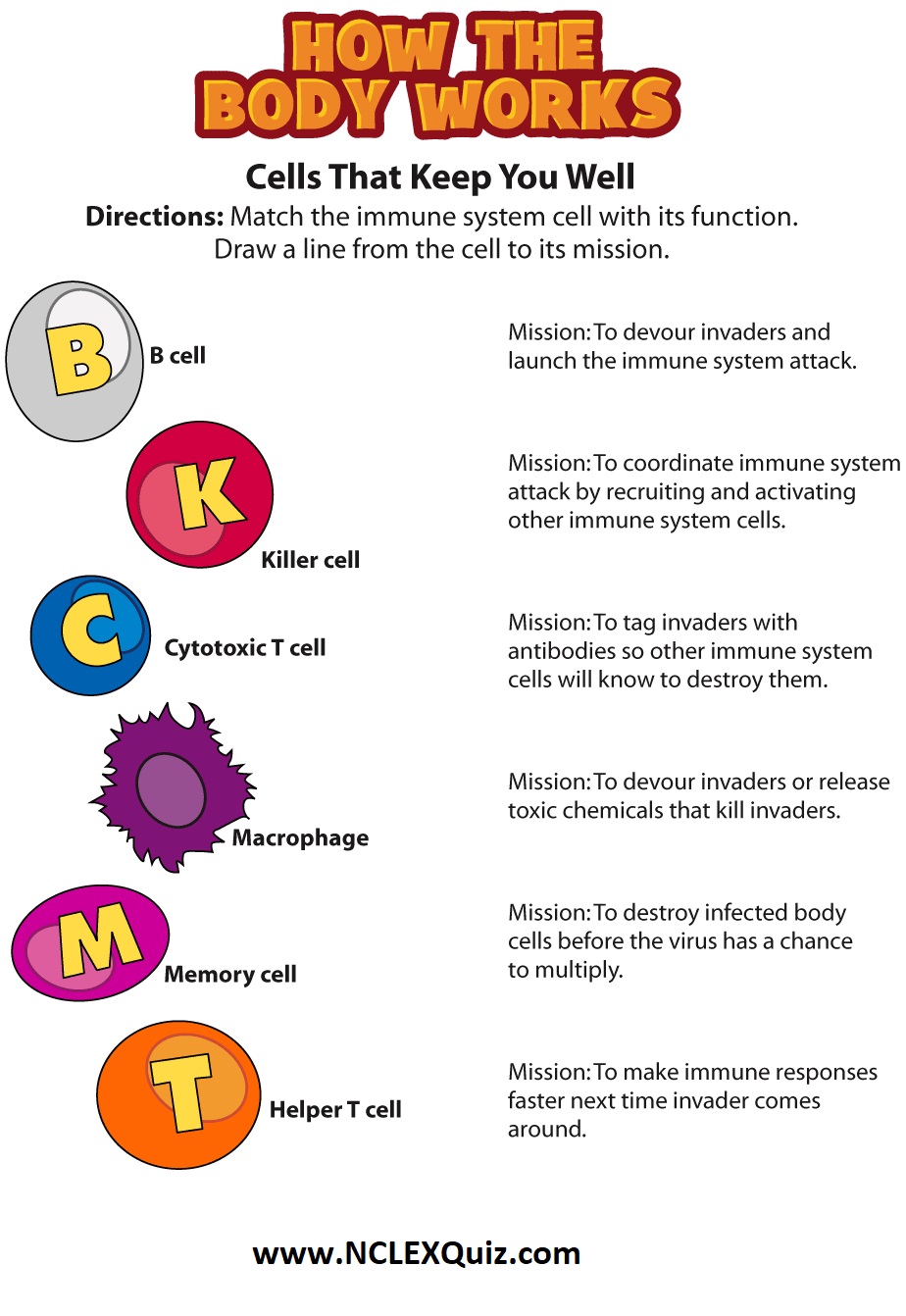
Do you know your immune system? Label the parts of the system that keeps you well.

What is the immune system?
The immune system protects your child’s body from outside invaders, such as bacteria, viruses, fungi, and toxins (chemicals produced by microbes). It is made up of different organs, cells, and proteins that work together.

There are two main parts of the immune system:

1. The innate immune system, which you are born with.
2. The adaptive immune system, which you develop when your body is exposed to microbes or chemicals released by microbes.

These two immune systems work together.

The innate immune system
This is your child’s rapid response system. It patrols your child’s body and is the first to respond when it finds an invader. The innate immune system is inherited and is active from the moment your child is born. When this system recognizes an invader, it goes into action immediately. The cells of this immune system surround and engulf the invader. The invader is killed inside the immune system cells. These cells are called phagocytes.

The acquired immune system
The acquired immune system, with help from the innate system, produces cells (antibodies) to protect your body from a specific invader. These antibodies are developed by cells called B lymphocytes after the body has been exposed to the invader. The antibodies stay in your child’s body. It can take several days for antibodies to develop. But after the first exposure, the immune system will recognize the invader and defend against it. The acquired immune system changes throughout your child’s life. Immunizations train your child’s immune system to make antibodies to protect him or her from harmful diseases.

The cells of both parts of the immune system are made in various organs of the body, including:

Adenoids. Two glands located at the back of the nasal passage.

Bone marrow. The soft, spongy tissue found in bone cavities.

Lymph nodes. Small organs shaped like beans, which are located throughout the body and connect via the lymphatic vessels.

Lymphatic vessels. A network of channels throughout the body that carries lymphocytes to the lymphoid organs and bloodstream.

Peyer’s patches. Lymphoid tissue in the small intestine.

Spleen. A fist-sized organ located in the abdominal cavity.

Thymus. Two lobes that join in front of the trachea behind the breastbone.

Tonsils. Two oval masses in the back of the throat.

Immune System Overview for Nursing Students (Structure and Function)

Placenta variations: Succenturiate Lobe and Velamentous

Placenta variations: Succenturiate Lobe and Velamentous

Velamentous Insertion: This is when some of the umbilical vessels run through the membranes (rather than attaching directly into the placenta). Although most babies are born fine, this kind of placenta does come with more risks. Because the vessels are unprotected, if the membranes (the bag of waters) is broken, it can puncture the vessels and cause hemorrhaging. Luckily, all the placenta’s that I have seen with this variation have all had happy endings.

Succenturiate Lobe: This is an extra lobe that is embedded in the membranes and connected to the main disc of the placenta via arteries/veins. According to radiopaedia.org this occurs in around 2/1000 pregnancies. It’s not really known what causes these accessory lobes. One theory put forth by babyworld.co.uk is that a slight irregularity or tiny scar on the uterine wall makes a small area inhospitable to the growing placenta, forcing a lobe to ‘move over’ and grow elsewhere. Some lobes are attached directly to the placenta via a ‘parenchymal bridge’, this is called a digitate lobe.

Placenta variations: Succenturiate Lobe and Velamentous

Placenta variations: Circummarginate and Circumvallate

Placenta variations: Circummarginate and Circumvallate

Circumvallate: This is when the fetal membranes create an edge of double folded membranes. It creates a thickened ring that makes a smaller circle (inwards from the edge of the placenta). The exact cause is unknown but midwife Patricia Edmonds says it can be caused by abnormal implantation of the placenta and the placenta and uterine wall growing at different rates.

Circummarginate: On this placenta, the point where the membranes attach is inside the edge of the placenta. The margin is thin and flat (whereas the circumvallate placenta has a thick ridge).

Placenta variations: Circummarginate and Circumvallate

Placenta variations: Bilobed & Battledore

Placenta variations: Bilobed & Battledore

Bi-lobed: This is when the placenta has two near equal-sized lobes. It may form if the uterus is an unusual shape. It can also be caused by one part of the placenta implanting in the front of the uterus and another part of the placenta attaching to the back of the uterus. It is NOT caused by a twin pregnancy (even though this is a common myth). According to radiopaedia.org it is estimated to occur in around 4% of pregnancies.

Battledore/Marginal Insertion: This is when the umbilical cord inserts into the rim of the placenta (rather than in the middle). It is called ‘battledore’ because the placenta looks like a racket used in the game battledore (similar to badminton). If the umbilical cord inserts not quite centrally and not quite on the margin, it is called an eccentric insertion.

Placenta variations: Bilobed & Battledore

Placenta variations and what they look like

Normally a human placenta is round like a plate, with a diameter of about 22cm and is about 2-2.5cm thick. It can weigh about 500 grams (and is usually heavier with bigger babies). It has two very distinct sides. The maternal side (the side attached to the mother’s uterus) is usually deep red/maroon and is made up of lobes (called cotyledons). The fetal side looks like a tree with veins and arteries running over the surface. The umbilical cord is usually 50-60cm long and has two arteries and a vein. It normally inserts into the middle of the placenta. Just like each baby is unique, each placenta has it’s own characteristics, and some have variations listed below.

Types of placenta previa
Partial. The placenta only partially covers the opening of the cervix. Vaginal birth is still possible.
Low-Lying. This type begins in early to mid pregnancy.
Marginal. The placenta begins to grow at the bottom of the uterus.
Major or complete. This is the most serious type.

Placenta variations and what they look like

Nursing Study Guide: Cellulitis

Cellulitis is an infection of the skin or underlying tissues.

Bacteria can be introduced into the skin through an area of open skin, such as an insect bite. However, in many cases, there is not an obvious site where bacteria entered the skin. Once bacteria are in the skin, they cause redness and swelling that can spread rapidly. Cellulitis can happen almost anywhere on the body but the most common place it occurs is the lower legs.

Symptoms of Cellulitis

Signs of cellulitis include redness of the skin (especially redness that spreads rapidly along the skin), warmth under the skin, and fever. The affected area can be painful. Certain bacteria can cause pus to collect beneath the skin (abscesses) or create blisters (bullae). Many different bacteria can cause cellulitis, but the most common are streptococci (especially beta-hemolytic streptococci) and Staphylococcus aureus.

Some patients are at increased risk of cellulitis, especially those with chronic swelling (lymphedema), those who are obese, and those with diabetes. Sometimes cellulitis can develop at the site of an animal bite or water exposure.

Diagnosis of Cellulitis

Cellulitis is diagnosed by physical examination. Laboratory testing and x-rays are not required. However, with more severe skin infections, studies like computed tomography scans and ultrasound might be done to look for deeper pockets of infection (abscesses).

Treatment

Cellulitis is treated with antibiotics to kill the bacteria involved. Antibiotics can be given by mouth (pills, liquid) in many cases, but in more severe cases, patients may need to be hospitalized for intravenous antibiotics. When an abscess occurs, it should be drained whenever possible. If a patient has frequent episodes of cellulitis, sometimes an antibiotic can be given daily for several weeks or even months to help prevent further episodes.

Preventing Cellulitis
  • Keep skin moisturized to prevent cracks and breaks in the skin, especially the feet.

  • Prevent formation of wounds.

  • Patients with chronic swelling should elevate the affected areas or use compression garments.

  • Take preventive antibiotics if prescribed by your doctor.

Nursing Study Guide: Cellulitis